

Imaging based diagnostic tools are pivotal in medical industry as they offer critical insights into several diseases accurately and facilitates rapid detection. Today, advancements in medical imaging enable healthcare professionals to visualize interior or structures that are not visible to naked eye and aid them in treatment planning and monitoring. Ophthalmology imaging has gained attention in recent times for its rapid development in ocular and orbital diseases evaluation.
Though ophthalmoscope is known to be one of the most popular medical devices since its invention, there have been several other discoveries and inventions in the field over the years. Emergence of various imaging techniques like fluorescence angiography, Scanning Laser Ophthalmoscopy (SLO) and Optical Coherence Tomography (OCT) in ophthalmology has benefited both healthcare professionals and patients in many ways from rapid diagnosis to timely patient care.
In this article, we look at one of the key diagnostic techniques in ophthalmology called retinal imaging. It requires a camera with sensitivity in the NIR (Near Infra-Red) spectrum to be able to capture clear images of the retina.
But before we learn how a camera solution with CMOS sensor plays a key role in imaging techniques in Ophthalmology, let us first try to understand what is retinal imaging.
Retinal imaging
Retinal imaging is a non-invasive imaging technique that produces high resolution images of the retina, blood vessels and the optic nerve. It is a replacement of the routine eye tests and helps ophthalmologists in more precise diagnosis of several ocular diseases and abnormalities including macular degeneration, glaucoma and diabetic retinopathy.
For instance, fundus photography is one such retinal imaging technique in ophthalmology and is used for diagnosis and documentation. Ophthalmologists rely on fundus cameras for swift examination of the retina and internal structures of the eye. There is a growing need for compact and portable fundus cameras that can provide high resolution images for near patient testing. Traditional fundus photography involves a procedure where the patient is given mydriatic eye drops which dilates the pupil. This can cause discomfort to the patient and is a time-consuming process. Today several non-mydriatic fundus imaging techniques have simplified the imaging process without the need for pupil dilation. Modern fundus cameras with high sensitivity and NIR imaging capabilities allow ophthalmologists to capture images of the retina without any loss of detail with less turnaround time.
Using NIR in retinal imaging
Around 4.1 million people at the age of 40 and above suffer diabetic retinopathy in the United States. NIR imaging is critical in early diagnosis of diabetic retinopathy and other ocular diseases like vascular occlusions, macular degeneration, and melanomas. NIR imaging is a non-invasive technique used to map pathological changes and detect alterations in the retinal pigments. This technique uses illumination which avoids back reflection from the cornea and captures the retina simultaneously.
For sub-retinal characteristics, near infrared light is more apparent than visible light. The optic nerve head, retinal vessels, and choroidal vessels seem dark, whereas sub-retinal deposits appear bright and thickened. As the wavelength is increased from 795 to 895 nm, the contrast and visibility of features rise. NIR fundus camera facilitates imaging of the interior of the human eye capturing multi spectral NIR images. Retinal imaging with spectral reflectance helps in identifying retinal conditions and provides more information about diabetic retinopathy, and melanomas making it suitable for fatal disease diagnosis.
Current trend in fundus retinal imaging is portable and affordable cameras with high quality sensors that facilitate high resolution output critical for accurate diagnosis.
High performance CMOS replacing CCD technology
A camera sensor is a critical component in ophthalmologic devices, and clinicians rely on high quality and high-resolution image data for better diagnosis and proposing suitable treatment plan.
Charge Coupled Device (CCD) and Complementary Metal Oxide Semiconductor (CMOS) are the two types of sensor technologies available in the market. CCD sensors are used in various scientific applications where light sensitive pixels are converted into signals. These high light sensitivity sensors capture images with low noise. Cameras with CCD sensors had been widely used for scientific imaging purposes and came with larger photodiodes for ophthalmologic imaging.
Today CMOS sensors are preferred over CCD sensors predominantly owing to their cost advantages. Earlier CMOS technology had a reputation for producing image outputs with noise. However, in addition to having a parallel A/D architecture to increase frame rates, correlated double sampling before as well as after each A/D – instead of just before the A/D – greatly improved SNR (Signal to Noise Ratio).
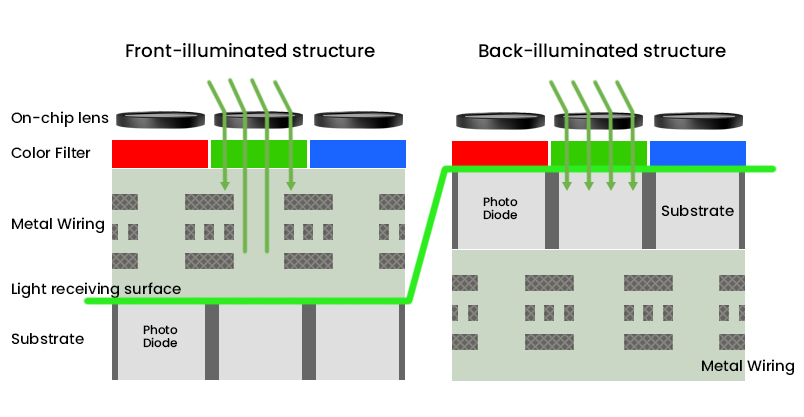
Also, CMOS sensors’ front-illuminated pixels which use metal wiring on top of the silicone that reflect some incoming photons and reduce the sensor’s sensitivity to light, are counter acted somewhat by using micro lenses. These micro lenses redirect incoming photons away from the wiring toward the photosensitive area of the pixel. To further improve sensitivity, CMOS technology then evolved from front-illumination to back-illumination. In back-illumination, the sensor is turned around so that the wiring and circuitry that can impede incoming photons is moved to the back side of the sensor and “out of the photons’ way”.
The following image illustrates the difference between front illuminated and back illuminated CMOS sensor technologies.
All these led to the improvement of image quality offered by CMOS sensors. Hence, they started becoming a natural choice over CCD sensors over time, especially for medical and life sciences applications.
IMX462 based See3CAM_CU27 for retinal imaging
e-con Systems™ has developed See3CAM_CU27, a Sony STARVIS IMX462 based USB3 camera for medical point of care devices. Being from the Sony STARVIS series, IMX462 sensor offers high sensitivity and greater picture quality in both visible light and near-IR regions.
See3CAM_CU27 is an ideal camera solution for various medical point of care and ophthalmological devices owing to 5 features
- High sensitivity
- Spectral bandwidth
- High frame rate
- Low noise
- Dynamic range
Let us now look at each of these in detail.
High sensitivity
See3CAM_CU27 offers high sensitivity with large pixels and produces high quality image output in low light conditions with its back side illuminated (BSI) pixels and low dark current. Having a high quantum efficiency helps to convert photons to electrons more efficiently, thereby capturing retinal images with less noise for accurate clinical observations.
Spectral bandwidth
Most ophthalmologic devices use NIR lighting for retinal imaging purposes. See3CAM_CU27 that houses IMX462 with high spectral bandwidth allows clinicians to capture superior NIR images of the structures of the retina.
This camera has high levels of quantum efficiency in the visible as well as the near infrared spectral regions. The photodiode portion of the pixel well is physically deeper compared to other BSI sensors, allowing photons of longer wavelength to penetrate deeper into the substrate. This dramatically increases the sensor’s sensitivity to red and NIR light. The camera sensor displays almost equal peak sensitivity to NIR light as it does to light in the visible spectrum. The peak QE (Quantum Efficiency) in the NIR around a wavelength of 800nm is as high as the peak QE in the visible wavelengths.
High framerate
IMX462 sensor based See3CAM_CU27 can deliver images at a high frame rate of more than 100fps. This is very desirable for ophthalmology imaging. This enables automated specimen scanning of retinal images and allows clinicians to diagnose diseases with accuracy.
Low noise
See3CAM_CU27 comes with SHCG (Super High Conversion Gain) for very low read noise at high gain. This is also helpful in improving SNR in low light conditions.
Dynamic range
The maximum number of electrons per pixel is known as the full well or saturation capacity. The dynamic range of a sensor is represented by the ratio of full well capacity to sensitivity threshold. The more the saturation capacity of a CMOS sensor, higher is the value of dynamic range. See3CAM_CU27 with IMX462 comes with high dynamic range and is best suited for retinal imaging owing to its ability to adapt to varying lighting conditions.
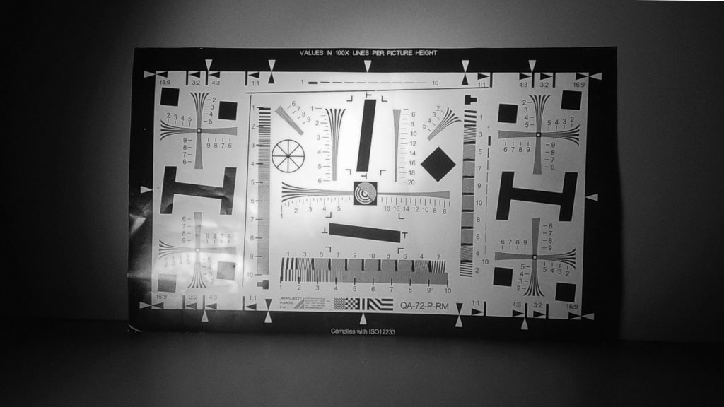
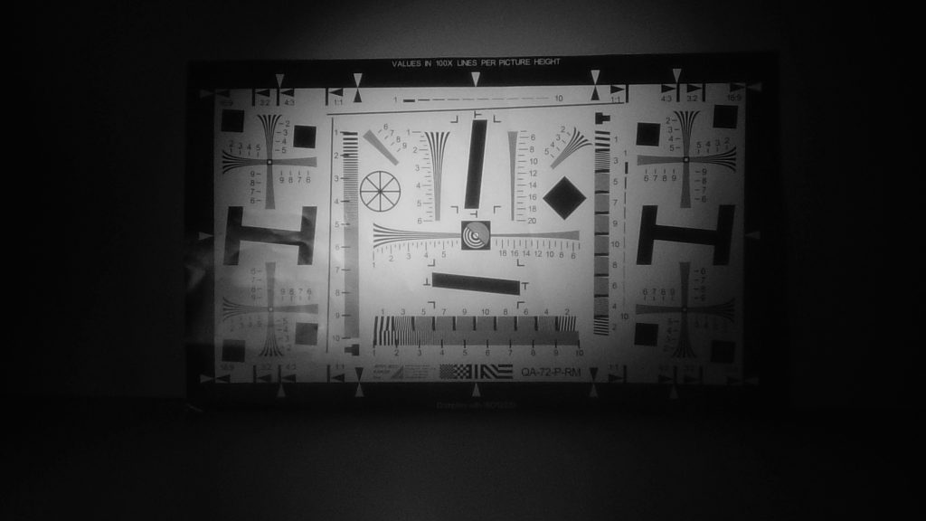
Comparison between CU27 and standard monochrome camera
Now that we have understood how See3CAM_CU27 is suitable for retinal imaging, let us learn how it compares with a normal monochrome camera.
Monochrome cameras, without their RGB filters are generally more sensitive to photons resulting in higher QE. But See3CAM_CU27’s NIR performance is much better, as it can be seen from the images below.


As explained in this blog, we can pretty much conclude that CMOS based vision systems are preferred for medical and life sciences applications, especially for ophthalmology. We also saw that See3CAM_CU27 is a perfect choice for retinal imaging. e-con Systems™ has already worked with a few customers where we helped them enhance the performance of their ophthalmology devices. To know more about this camera solution, visit the See3CAM_CU27 product page.
Related Products:
1. See3CAM_CU27 – Sony® Starvis™ IMX462 based ultra-low light USB 3.1 Gen 1 Camera
2. e-CAM22_CUNX – Sony® Starvis™ IMX462 ultra-low light camera for NVIDIA Jetson Xavier NX platform
3. e-CAM23_CUXVR – Sony® Starvis™ IMX462 ultra-low light camera for NVIDIA Jetson AGX Xavier platform

Vinoth Rajagopalan is an embedded vision expert with 15+ years of experience in product engineering management, R&D, and technical consultations. He has been responsible for many success stories in e-con Systems – from pre-sales and product conceptualization to launch and support. Having started his career as a software engineer, he currently leads a world-class team to handle major product development initiatives