

Blood smear testing has been gaining popularity for its accurate diagnosis of various blood disorders. Medical practitioners usually suggest a peripheral blood smear test as part of the routine complete blood count test.
In this blog, you’ll get to learn more about this testing process and the significance of embedded camera solutions in blood smear testing.
A Brief Overview of Blood Smear Testing
A blood smear test involves spreading a blood sample on a microscopic slide treated with a special stain. These stained slides are then examined under digital microscopes with cameras mounted on tops.
A blood smear helps us analyze the size, shape, and other characteristics of blood components. It is an effective visualization tool, helping recognize abnormalities in blood cell morphology that go undetected in the usual CBC tests. Changes in the size or shape of blood components detected by a blood smear test could help diagnose various underlying blood disorders.
Let’s now see what a blood smear test helps detect:
Size of each blood cell type: Every blood cell must be within a specific size range. Any deviation from the specified range could indicate conditions like Macrocytic Anaemia and Microcytosis.
- Shapes of various types of blood cells: A blood smear test helps detect conditions like sickle cell disease and elliptocytosis, which are cases of abnormally shaped red blood cells.
- Color variations in RBCs: The intensity of red color in the blood directly correlates with its hemoglobin content, which in turn determines the blood’s oxygen-carrying capacity. Any alterations in the recommended blood cell hues can be readily identified through a blood smear examination.
- The presence of NRBCs: Nucleated Red Blood Cells in adult blood is uncommon. They could indicate various disorders, like leukemia or hypoxia (reduced oxygen in the blood). A blood smear test is done to confirm the presence of NRBCs.
- Abnormalities in WBC differentials: WBCs, comprising lymphocytes, monocytes, neutrophils, eosinophils, and basophils, can be challenging to discern due to their similar size and structure. However, a blood smear test aids in detecting shape or size abnormalities, enabling accurate diagnosis.
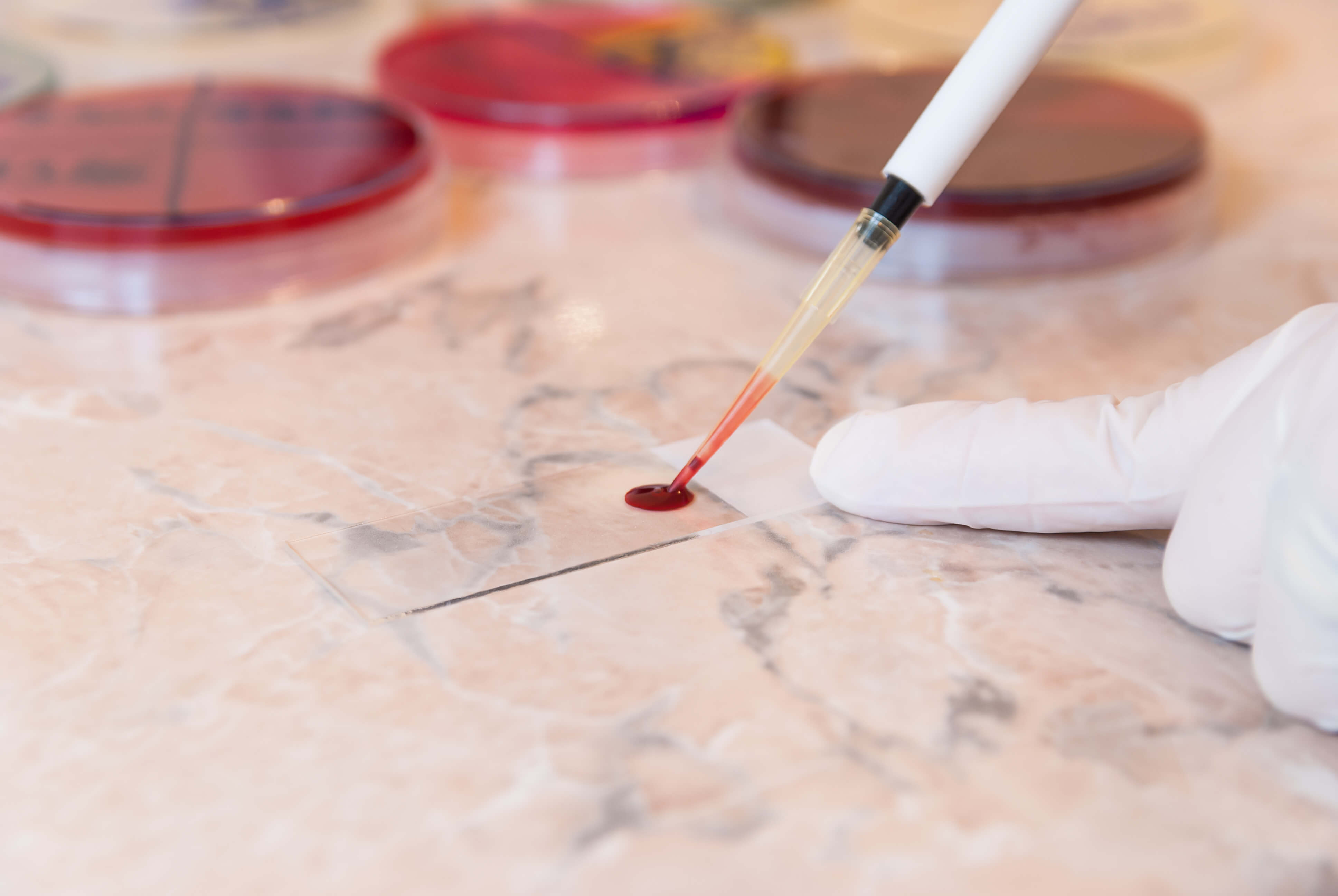 Image: A sample of Blood Smear Technique
Camera Features that Overcome the Blood Smear Testing Challenges
The entire process of diagnosis in blood smear testing is linked to the quality of the images produced. The clarity and accuracy of these images directly influence diagnostic assessments. High-resolution images are essential for accurate interpretation by healthcare professionals and for remote consulting scenarios.
Let’s now examine the common challenges faced by the medical community with blood smear testing and how the right camera solutions help elevate them.
Accurate color reproduction of stains in the blood smear image
Blood smear slides are usually stained with stains like Leishman stain and Wright-Giemsa. Staining is done to enhance the contrast as well as the distinct characteristics of the different types of blood cells. The below-mentioned features ensure accurate color reproduction in Blood Smear Imaging.
- Spectral sensitivity: It refers to a sensor’s responsiveness to the various colours in the visible and near-infrared spectrums of light. Cameras with sensors of well-calibrated spectral sensitivity help in capturing the colours in the image.
- High-quality inbuilt processing: Inbuilt processing systems play a crucial role in color calibration and white balance adjustments. It enhances the contrast of the image, ensuring the colours are reproduced accurately.
Minimization of imaging artifacts
The usage of low-quality cameras in blood smear testing can result in various artifacts in the final image causing a significant amount of negative impact. These imaging artifacts could be pixelation, temporal noise, photobleaching and many more. Let’s now explore a few imaging artifacts and the camera features that help mitigate them.
- Pixelation: These artifacts occur when the magnification level exceeds the image sensor’s capability, resulting in a visible pixel boundary that degrades and reduces spatial resolution. To resolve pixelation, cameras can apply Shannon’s sampling theorem, ensuring that the sampling interval is no greater than one-half the size of the smallest resolvable feature, thereby capturing sufficient detail without over-sampling.
- Temporal noise: It usually arises from spatial and temporal variations when subjected to constant uniform illumination. This artifact increases the image graininess, resulting in a loss of detail. It can be minimized using exposure time adjustments and high SNR cameras.
- Photobleaching: It occurs when organic molecules like blood cells absorb photons at visible wavelengths, leading to chemical reactions that render these cells non-functional. To mitigate this artifact, illumination control measures and enhancing the detection efficiency through factors like numerical aperture and global shutter can be done.
Therefore, the camera must be equipped with advanced noise reduction algorithms and a high-quality sensor to ensure crystal-clear images even in low-light conditions.
Overcoming the challenge of cell differentiation
Blood cell differentiation in blood smear testing poses a major challenge due to their similar appearances under the microscope. It can lead to misidentification of cells, impacting diagnostic accuracy and patient care. Hence, it is imperative to utilize high-resolution sensor-based cameras that can capture fine details.
High-resolution cameras offer enhanced definition, making it easy to accurately detect subtle differences in cell morphology. With a greater number of pixels, high-resolution cameras can capture finer details and nuances in blood smear images.
Cameras with a high dynamic range ensure that cellular features stand out distinctly against the background. So it becomes simpler to distinguish between different cell types and abnormalities. Enhanced contrast also enhances image clarity.
Limitations of a slower processing time
In blood smear imaging, there’s a need to examine a larger area of the blood smear efficiently. This broader examination allows for the observation of a greater number of blood cells. However, the conventional approach of capturing multiple images and stitching them together can be time-consuming, hindering the diagnosis.
Imaging systems with a wider field of view offer a solution by allowing the capture of a larger area of the blood smear in a single image. Systems with a wider field of view enhance workflow efficiency and enable faster processing of blood smear samples by streamlining the imaging process.
The utilization of wider field-of-view imaging systems also improves diagnostic accuracy, enabling the evaluation of a greater number of blood cells in context.
World-class Medical Cameras Offered by e-con Systems
e-con Systems, with over two decades of experience in designing, developing, and manufacturing OEM cameras, offers a wide range of imaging solutions to build cutting-edge medical devices.
Our Sony Pregius Camera series is designed with the best CMOS image sensors with best-fit features ideal for applications in the areas of peripheral blood smear testing, point of care, fundus imaging, remote patient monitoring, surgery, etc.
Browse the Camera Selector Page to see our full portfolio.
If you want to get started integrating our cameras into your medical products, please write to camerasolutions@e-consystems.com.

Balaji is a camera expert with 18+ years of experience in embedded product design, camera solutions, and product development. In e-con Systems, he has built numerous camera solutions in the field of ophthalmology, laboratory equipment, dentistry, assistive technology, dermatology, and more. He has played an integral part in helping many customers build their products by integrating the right vision technology into them.