

Photomicrography is vital in medical and life sciences, enabling scientists and researchers to capture and analyze microscopic images with exceptional clarity and detail. This blog will explore the key components of photomicrography, its significance in various applications, and the challenges associated with camera technology. Furthermore, we will delve into how the See3CAM_50CUG camera from e-con Systems addresses these challenges and revolutionizes the capabilities of devices that utilize photomicrography.
What is Photomicrography?
Photomicrography is the technique of capturing high-resolution images of microscopic specimens using specialized cameras mounted on microscopes. It enables visualization and analysis of intricate details, aiding in scientific research, medical diagnostics, and several life science applications.
Key Components in Photomicrography
The key components of photomicrography include a microscope with suitable optics, lighting systems for illumination, camera systems, and image capture and processing software. Each component contributes to the overall image quality and accuracy.
- Microscope with suitable optics: The microscope is the primary tool in photomicrography. It comprises various optical components, including lenses, objectives, eyepieces, and condensers, designed to magnify and resolve microscopic specimens.
- Lighting systems for illumination: Proper illumination is crucial in photomicrography to enhance the visibility and contrast of microscopic specimens. Lighting systems, such as brightfield, darkfield, phase contrast, or fluorescence illumination, are employed.
- Camera systems: Camera systems are integral to photomicrography as they capture the magnified images produced by the microscope. Dedicated microscopy cameras are designed to provide high-resolution imaging, low noise, and excellent sensitivity to capture fine details.
- Image capture and processing software: Once the camera captures the image, it is processed and stored using specialized software. Image capture software enables users to control camera settings, adjust exposure parameters, and acquire images with precise control.
The Role of Photomicrography in Medical and Life Sciences
- Pathology and histology: Photomicrography is utilized to analyze tissue samples for disease diagnosis and research in pathology and histology. By capturing detailed images of cells and tissues under a microscope, pathologists can identify abnormalities, study disease progression, etc.
- Microbiology: Microbiologists employ photomicrography to study microorganisms, such as bacteria, viruses, and fungi. These images allow researchers to observe their behavior, interactions, and structural characteristics.
- Genetics and cytogenetics: Photomicrography plays a crucial role in genetics and cytogenetics by enabling researchers to examine chromosomes and genetic materials. By capturing detailed images of chromosomes, scientists can study genetic abnormalities, chromosomal rearrangements, and gene expressions.
- Cell biology: Photomicrography is extensively used in cell biology to investigate cellular processes, organelles, and molecular interactions. Researchers can visualize and analyze cellular phenomena by capturing images of cells and cellular structures, such as cell division, protein localization, intracellular trafficking, and signal transduction pathways.
- Neurobiology: In neurobiology, photomicrography is instrumental in visualizing neural structures and studying brain function. By capturing detailed images of neurons, synapses, and neural circuits, researchers can explore the intricacies of the nervous system, map neuronal connections, and investigate the underlying mechanisms of brain disorders.
- Developmental biology: Photomicrography is widely utilized in developmental biology to observe embryonic development and tissue morphogenesis. By capturing sequential images of developing embryos and tissues, researchers can analyze the processes involved in organ formation, cell differentiation, and tissue patterning.
- Pharmaceutical research: In pharmaceutical research, photomicrography is used to assess the effects of drugs on cells and tissues. Researchers can evaluate drug efficacy, cytotoxicity, and potential side effects by capturing images of cells treated with different compounds. These images provide visual evidence of the drug’s impact at a microscopic level and aid in developing safe and effective therapeutic interventions.
Camera-Related Challenges in Photomicrography
Photomicrography has its fair share of challenges, particularly regarding camera equipment and techniques. These challenges need to be addressed to ensure high-quality and accurate results.
Resolution and image quality
Resolution and image quality are crucial factors in photomicrography. The ability to capture fine details and maintain image fidelity is essential for accurate analysis and interpretation. Microscopic subjects often have intricate structures, and capturing them with clarity requires high-resolution cameras with advanced optics. Additionally, ensuring proper alignment and focus is essential to minimize any aberrations or distortions that could compromise the image quality.
Low-light sensitivity
Low-light sensitivity is another significant challenge in photomicrography. Many microscopic subjects require specific lighting conditions, such as fluorescence or darkfield illumination. Obtaining clear images in such low-light conditions can be challenging, especially without introducing excessive noise or compromising the image quality. Using cameras with high ISO capabilities, low-noise sensors, and proper lighting techniques can help overcome this challenge.
Speed and responsiveness
Speed and responsiveness are critical in capturing fast-moving samples or dynamic processes in photomicrography. Microscopic subjects often exhibit rapid movements or changes, and capturing them without motion blur requires cameras with fast shutter speeds and high frame rates. Additionally, specialized techniques like high-speed imaging or time-lapse photography can help freeze the motion and effectively capture the desired details.
Color accuracy
Color accuracy is particularly important in photomicrography, especially with fluorescent imaging. Fluorescent dyes and stains commonly label specific structures or molecules in microscopic samples. However, capturing and reproducing accurate colors can be challenging due to the complex nature of fluorescence and the variations in color response among different cameras and imaging systems. Calibrating the camera settings and using color correction techniques can ensure a faithful representation of colors in photomicrographs.
Data transfer and storage
Managing large volumes of high-resolution image data is a significant challenge in photomicrography. Microscopic imaging often generates a vast amount of data, especially when capturing images at high resolutions or using multi-channel imaging techniques. Storing, organizing, and transferring large data sets require robust data management solutions, including high-capacity storage devices, efficient data transfer protocols, and well-organized file systems. Implementing proper backup and archiving strategies is crucial to prevent data loss and ensure long-term accessibility.
How e-con Systems’ See3CAM_50CUG Addresses These Challenges
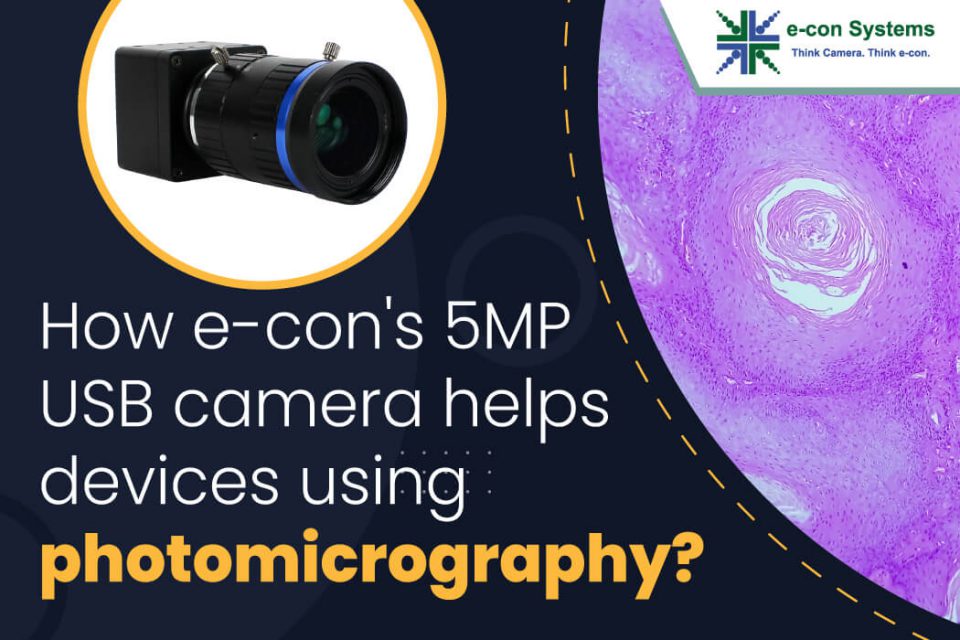
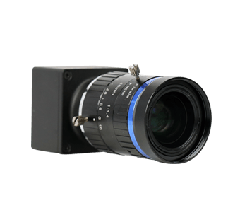
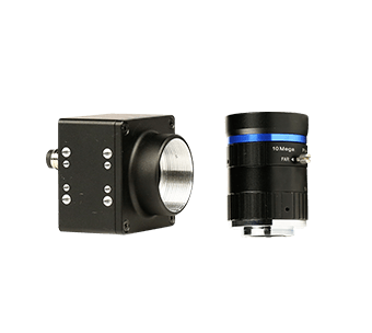
The See3CAM_50CUG is a 5MP USB camera developed by e-con Systems that presents a breakthrough solution for enhancing photomicrography. It offers unparalleled imaging capabilities with key features such as:
- High-resolution imaging: The 5MP Sony Pregius IMX264 sensor provides exceptional detail and sharpness.
- Global shutter technology: Eliminates motion artifacts, enabling precise imaging of moving samples.
- Excellent low-light performance: The camera’s advanced sensor and noise reduction capabilities ensure clear images even in challenging lighting conditions.
- Color accuracy: The color variant of See3CAM_50CUG accurately captures and reproduces vibrant colors in fluorescence imaging.
- USB interface: Offers convenient connectivity and high-speed data transfer.
- Comprehensive software support: The camera has various software tools and SDKs, enabling seamless integration and control.
We can expertly customize your camera parameters to ensure the best possible image quality under custom light conditions – specific to your application. Our fine-tuning expertise also makes us a standout embedded vision partner of choice.
So, if you want to integrate high-performance cameras into your photomicrography applications, please write to us at camerasolutions@e-consystems.com.
You can also explore all our medical and life science camera solutions or check out our full portfolio.

Balaji is a camera expert with 18+ years of experience in embedded product design, camera solutions, and product development. In e-con Systems, he has built numerous camera solutions in the field of ophthalmology, laboratory equipment, dentistry, assistive technology, dermatology, and more. He has played an integral part in helping many customers build their products by integrating the right vision technology into them.