

Fundus photography offers a deeper look into the back of the eye, known as the fundus or retina. It provides a detailed view of the fundus region, helping with diagnosis, monitoring, and management of various ophthalmic diseases. It also enables patient education by empowering ophthalmologists to explain the condition and its’ progression.
Fundus photography is becoming increasingly prevalent, given the widespread chronic eye disorders like diabetic retinopathy and age-related macular degeneration, especially among the elderly. That’s why a recent report has stated that fundus cameras are expected to grow at a CAGR of 6.2% from 2023 to 2030.
In this blog, we’ll explore the technology behind fundus cameras, their types, modes, applications, future scope, and more.
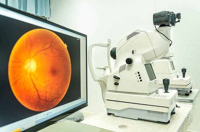
Image 1: A table-top fundus camera in use
How Do Fundus Cameras Work?
Fundus cameras utilize a controlled light source to illuminate the retina, capturing high-resolution images that allow for detailed visualization of the posterior segment, especially areas not easily visible with direct ophthalmoscopy. Light is directed into the eye through the pupil and passes through key refractive media—namely, the cornea, lens, and vitreous humor—before it reaches the retina.
The reflected light from the retinal surface is then focused back through these structures and imaged onto the sensor (either CCD or CMOS) within the camera system.
The optical design of the camera often uses multiple lenses and filters, which ensures precise magnification by enhancing specific retinal features. Filters like red-free or infrared can be applied to isolate vascular structures, choroidal layers, or pathological changes, depending on the clinical needs.
What Are the Types of Fundus Cameras?
Fundus cameras are categorized based on factors like pupil dilation requirements, FoV, stereoscopic capabilities, clinical applications, etc. Let’s look at the five types of fundus cameras:
- Mydriatic cameras
- Non-mydriatic cameras
- Stereoscopic cameras
- Scanning laser ophthalmoscope (SLO)
- Ultra-widefield cameras
1. Mydriatic fundus cameras
These cameras require pharmacological dilation of the pupil (typically using tropicamide) to enhance the field of view and illumination quality. They utilize a high-intensity xenon or LED flash to capture images of the retina through a series of optical lenses and filters, producing high-resolution images (often 50° to 60° FOV). These systems play an integral role in diagnosing conditions like diabetic retinopathy, age-related macular degeneration, and other retinal diseases that need high contrast and thorough visualization of the posterior segment.
2. Non-mydriatic fundus cameras
These cameras operate without the need for pupil dilation, typically achieving a field of view (FOV) of 30° to 50°. They utilize infrared pre-imaging to align and focus before capturing the final image using a visible white light flash. The integration of near-infrared light sources (700-900 nm) allows the camera to bypass smaller pupils and minimize patient discomfort. Non-mydriatic cameras are widely used in mass-screening programs and routine ophthalmic examinations for conditions like glaucoma and hypertensive retinopathy.
3. Stereoscopic fundus cameras
Stereoscopic imaging uses dual optical paths to capture images from two slightly different perspectives. This allows for the reconstruction of 3D images of the retina. The system typically employs beam-splitting optics, capturing simultaneous or sequential stereo images to create depth perception, which is essential for evaluating retinal surface changes, such as edema and epiretinal membranes. This 3D visualization aids in assessing more complex retinal pathologies.
4. Scanning Laser Ophthalmoscope (SLO)
SLO technology replaces traditional flash illumination with laser scanning systems. These cameras use low-power lasers (often multi-wavelength: green 532 nm, red 633 nm, and infrared 780 nm) to scan the retina point-by-point. The confocal design improves image resolution and depth of focus, allowing imaging through small pupils (down to 2 mm). SLO is ideal for real-time imaging, higher contrast, and diagnostic applications where fine details are required, especially in teleophthalmology and peripheral pathologies.
5. Ultra-widefield fundus cameras
These systems capture a much larger retinal area in a single image, often up to 200° FOV, compared to the standard 45°-50° of traditional cameras. They often use specialized optics, including aspheric or ellipsoidal lenses, in combination with mirrors to broaden the field of view. Ultra-widefield systems can capture images in a single shot, making them indispensable for evaluating peripheral retinal conditions that include retinal tears, detachment, and peripheral ischemia in conditions like diabetic retinopathy.
Modes of Fundus Photography
Fundus photography modes are classified based on the light source (visible or near-infrared) and specific filters used to enhance different structures of the retina and underlying layers.
Color fundus photography
This method captures clear color images of the retina using white light and color filters, offering a real-time view of the posterior eye segment.
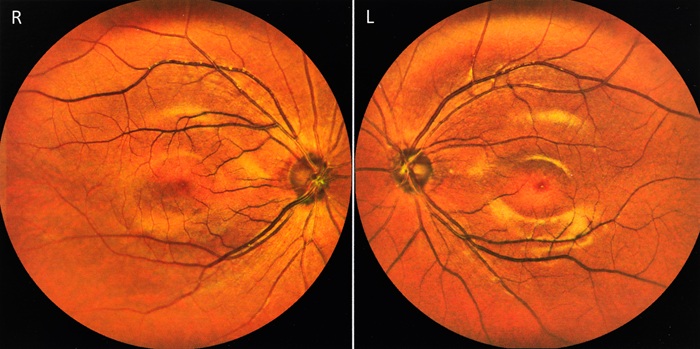
Image 2: An image of the human retina taken by a fundus camera
Red-free fundus photography
It employs a green filter (~540-570 nm) that blocks red light, improving contrast to highlight retinal blood vessels and lesions. This is often used as a baseline before other angiography techniques.
Fluorescein Angiography (FA)
This mode involves injecting fluorescein dye, which travels to retinal vessels –illuminated by blue light (~490 nm) to assess retinal vascular diseases.
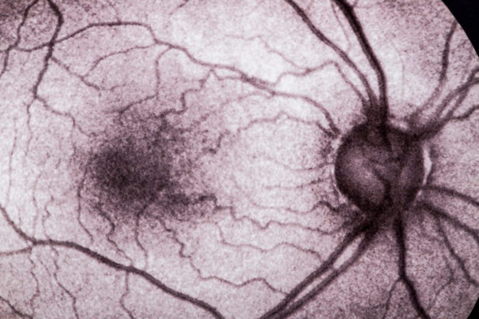
Image 3: An eye angiogram
Indocyanine Green Angiography (ICGA)
This mode uses indocyanine green dye and near-infrared light (500-810 nm) to visualize deeper choroidal blood vessels beneath the retina.
Popular Applications of Fundus Cameras
- Diagnosing and monitoring the damage to the blood vessels of the retina caused by diabetes, which is known as diabetic retinopathy.
- Identifying changes in the optic nerve and retina that might indicate glaucoma.
- Evaluating the progression of AMD and assessing the effectiveness of treatments.
- Detecting signs of retinal detachment or tears in the retina.
- Evaluating the retina before and after ocular surgeries.
- Collecting data for research on retinal diseases and testing new treatments.
- Providing a comprehensive evaluation of retinal health as part of regular eye check-ups.
Emerging Innovations in Fundus Photography
- AI and ML: Fundus Photography is no exception when it comes to the wave of AI/ML integration. Recent research has shown that AI-based DL algorithms for optic nerve disease performed well in differentiating glaucoma from non-glaucoma on color fundus photographs. AI-based retinal imaging tools are being adopted in clinical settings as well.
- Combination with OCT: Optical Coherence Tomography refers to a non-invasive imaging approach that offers high-resolution cross-sectional visuals of the retinal layers beneath the surface. Fundus photography, on the other hand, provides detailed images of the surface. Together, they complement each other in providing a complete picture of the retinal health.
- Augmented and Virtual Reality: Modern-day surgical procedures are adopting AR and VR systems into their workflows to enhance the surgeon’s vision. Fundus photographs could be overlaid with the surgeon’s field of view to guide surgical instruments with greater precision in AR-assisted retinal surgeries. These photographs can be used to create 3D models that support planning complex retinal surgeries.
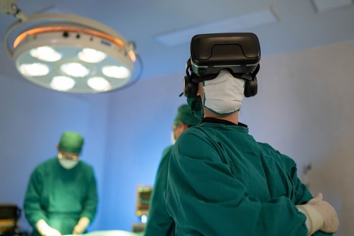
Image 4: The use of virtual reality glasses in surgeries
- Multispectral imaging: This imaging method involves the capture of image data at different wavelengths across the electromagnetic spectrum beyond the visible light range. When combined with fundus photography, it captures retinal images at different wavelengths. These include infrared or UV, which can reveal additional details about the retina that are not visible in standard fundus photography.
High-Performance Fundus Cameras by e-con Systems
Since 2003, e-con Systems has been designing, developing, and manufacturing OEM cameras. Over the years, we have equipped many clients with state-of-the-art camera solutions for their developers to create world-class fundus photography devices.
Check out our camera solutions for Fundus and Retinal Imaging.
e-con Systems also offers cameras that are perfect for other medical use cases such as dentistry, Point of Care, remote patient monitoring, surgery, etc. They come with features such as high QE, superior NIR performance, IP-rated enclosures, compatibility with embedded processors such as NVIDIA, NXP, Qualcomm, and Rockchip, and more.
See all our medical and life sciences cameras
Visit our Camera Selector Page to browse through our complete portfolio.
If you require an expert to integrate the right camera into your dental product, please write to camerasolutions@e-consystems.com.

Balaji is a camera expert with 18+ years of experience in embedded product design, camera solutions, and product development. In e-con Systems, he has built numerous camera solutions in the field of ophthalmology, laboratory equipment, dentistry, assistive technology, dermatology, and more. He has played an integral part in helping many customers build their products by integrating the right vision technology into them.