

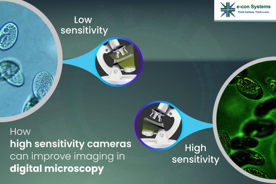
Digital microscopy has revolutionized the way we observe and analyze the microscopic world. It has become an indispensable tool in various medical and life science fields. With digital microscopy, images are captured and analyzed with the help of computer software, making it possible to obtain more detailed information and precise measurements than traditional microscopy methods. However, the quality of the captured images highly depends on the camera’s sensitivity.
In this article, you can explore how high sensitivity cameras improve imaging in digital microscopy, their clear-cut advantages, and some of the major use cases involved. You’ll also find out the latest details about our brand-new all-in-one camera – the See3CAM_50CUGM – a perfect fit for your digital microscope or any other medical and life science application.
What are high sensitivity cameras?
As you may have guessed (or known), high sensitivity camera solutions are capable of producing high-quality images even in situations where traditional solutions struggle. These cameras use advanced technologies such as larger sensors and better noise reduction to achieve exceptional sensitivity. This makes high sensitivity cameras ideal for capturing images in low-light conditions or in situations where fast shutter speeds are not possible.
Why high sensitivity cameras are preferred in digital microscopy
High sensitivity cameras have the ability to detect and capture low levels of light, which is crucial in many digital microscopy applications. In traditional microscopy, the amount of light that reaches the detector is limited by the optics of the microscope and the sensitivity of the camera. With the development of high sensitivity cameras, it is now possible to capture images of specimens that were previously too dim to be observed.
Advantages of high sensitivity cameras in digital microscopy
- Low light sensitivity: Digital microscopy often involves imaging very small samples or specimens that are illuminated with low-intensity light sources such as LEDs or lasers. High sensitivity cameras are capable of detecting even the faintest light signals, resulting in clearer and brighter images.
- Higher signal-to-noise ratio: High sensitivity cameras have a better signal-to-noise ratio, which means that the image is less affected by background noise or interference. It results in clearer images with better contrast and less noise.
- Improved dynamic range: High sensitivity cameras have a wider dynamic range, meaning they can capture a greater range of light intensities, from the darkest to the brightest parts of the sample. This helps to avoid over-exposed or under-exposed areas in the image.
- Faster image acquisition: High sensitivity cameras often have faster readout rates, allowing for faster image acquisition and processing.
Camera solution for Medical Microscope to Improve the Accuracy & Speed of Diagnosis
Use cases of high sensitivity cameras in digital microscopy
Fluorescence microscopy
Fluorescence microscopy is a technique that uses fluorescent dyes to label specific molecules in a specimen. When the specimen is illuminated with a specific wavelength of light, the labeled molecules emit light at a different wavelength. However, the emission of light is often very weak, making it difficult to capture a clear image. So, with high sensitivity cameras, it becomes seamless to capture these weak emissions.
Super-resolution microscopy
Super-resolution microscopy is a technique that allows for the imaging of structures that are smaller than the diffraction limit of light. In this case, the switching of the fluorescent dyes is often accompanied by a decrease in fluorescence intensity. So, it becomes challenging to capture clear images. But with high sensitivity cameras, you can capture the weak emissions, enabling the imaging of structures with higher resolution.
Live cell imaging
High sensitivity cameras can capture high-quality images of live cells without damaging them. They can also capture images at high frame rates – allowing the real-time capture of fast-moving processes. They also produce high-quality images for quantitative analysis of cellular processes.
Read More: Why global shutter cameras are a perfect fit for live cell imaging systems
Darkfield microscopy
High sensitivity cameras can capture clear images of specimens under darkfield illumination, making them useful for studying transparent or low-contrast samples. Also, such cameras can capture images of specimens labeled with different fluorophores simultaneously. So, it becomes possible to study multiple biological processes in a single experiment.
Why See3CAM_50CUGM, our latest release, is picture-perfect for digital microscopes
e-con Systems already has a proven track record in helping design custom camera solutions for medical and life science applications, including digital microscopes. Our latest release – the See3CAM_50CUGM – is a 5MP high-sensitivity fixed focus camera based on the Sony Pregius IMX264 CMOS sensor. It is the ultimate all-in-one camera for the medical and life science industry as it can be easily integrated into a wide range of applications – adapting to different imaging modes and techniques. Some of its differentiating features include:
- Global shutter efficiency
- High signal-to-noise ratio
- High dynamic range
- Short and long exposure periods
- Back-illuminated pixel technology
- Large pixel size of 3.45µm
- Peak quantum efficiency in the IR spectrum
- Trigger function for synchronization
- C-mount lens holder
- UVC compliant for Windows and Linux
Discover more about See3CAM_50CUGM
Check out our Camera Selector page
If you need help integrating camera solutions into your digital microscope or any other camera-based life science application, please write to us at camerasolutions@e-consystems.com.

Balaji is a camera expert with 18+ years of experience in embedded product design, camera solutions, and product development. In e-con Systems, he has built numerous camera solutions in the field of ophthalmology, laboratory equipment, dentistry, assistive technology, dermatology, and more. He has played an integral part in helping many customers build their products by integrating the right vision technology into them.