

Flow cytometers are basically instruments used to analyze and quantify the physical and chemical characteristics of individual cells or particles in a fluid stream. Embedded vision technology plays an important role in these flow cytometers by enabling the acquisition, analysis, and interpretation of large amounts of cellular data in real-time.
In this blog, you’ll find out more about what flow cytometers can do, how they work, and the role of embedded vision technology in their evolution.
What is a flow cytometer?
Flow cytometry is a common analytical method for counting, inspecting, and classifying cells floating in a stream of fluid. Flow cytometry is a technology that rapidly analyzes multiple physical characteristics of single cells or particles as they flow in suspension through a measuring device. Then, the cells are analyzed and differentiated using physical characteristics like size, granularity, and fluorescent features.
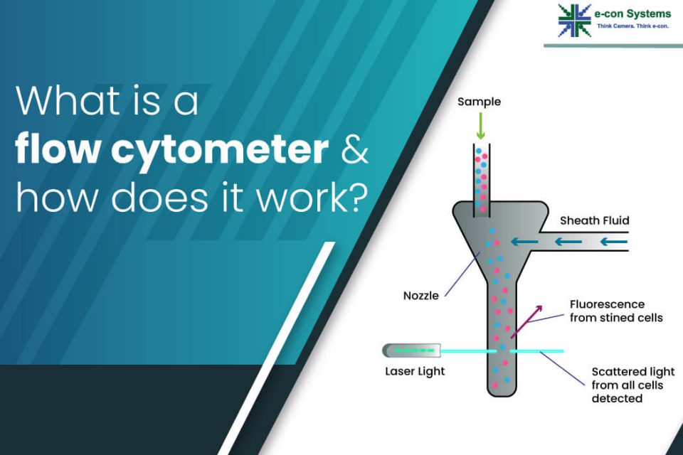
All flow cytometric techniques include the two fundamental phases of cell concentrating and detection. The cells must first be focused into a single file arrangement within the center of fluid flow to make the cells pass through the focal plane of a detection system. That way, only one cell is ever present in the detection volume. This method makes it possible to examine cells of interest in series using their optical (fluorescence emission or scatter) characteristic. An important benefit of the method is that it permits quantitative, non-destructive analysis of size, morphology (such as shape and internal complexity), and biochemistry (such as cell cycle distribution and DNA content).
Working principle of a flow cytometer: How it works
Flow cytometry follows a method that uses light scattering and fluorescence emission to study the movement of particles. Cell components are labeled with fluorescent material and then excited by a laser to emit light at different wavelengths. After that, the fluorescence can be analyzed to determine the number and kind of cells in a sample. Hence, it is possible to analyze up to a thousand particles each second as they move through the liquid stream.
A laser beam targets a hydrodynamically concentrated stream of fluid carrying the cells. Several detectors are carefully placed around the stream at the point where the fluid passes through the light beam to measure the light scattering. One detector is positioned parallel to the light beam to measure forward scatter, while another is perpendicular to the stream to measure side scatter.
When cells labeled with fluorescent are passed through the laser light, they are excited by the laser and emit light of a longer wavelength. Additional fluorescent detectors are placed to pick up this fluorescent light along with scattered light. All the detected signals are analyzed by a computer using specialized software to measure various information on cells’ physical and chemical structure. For instance, the forward scatter can detect cellular volume information, whereas the side scatter can detect inner cellular complexity.
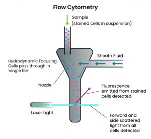 Figure 1. Overview of the flow cytometer.
Major use cases of a flow cytometer: Where it can be used
Flow cytometers can be used in various applications, including DNA sequencing, T-cell phenotyping, and detecting uncommon cells. Some of these use cases include:
- Immuno-phenotyping: Identify and characterize different types of immune cells, such as T cells, B cells, and natural killer cells, based on their surface markers.
- Cell cycle analysis: Analyze the distribution of cells in different phases of the cell cycle, providing insight into cell proliferation and DNA replication.
- Apoptosis analysis: Analyze the percentage of cells undergoing programmed cell death, or apoptosis, based on the binding of fluorescent dyes to DNA.
- DNA content analysis: Analyze the DNA content of cells, providing information on ploidy, chromosomal abnormalities, and cell proliferation.
- Stem cell analysis: Detect and isolate stem cells based on their surface markers, which can be used for regenerative medicine and tissue engineering.
- Cancer research: Analyze cancer cells and identify specific biomarkers that may be used for diagnosis, prognosis, and treatment.
- Microbiology: Quantify and analyze bacteria, viruses, and other microorganisms in environmental samples, such as water or soil.
- Drug discovery: Screen potential drug candidates based on their effects on cell signaling pathways and cellular processes.
How embedded vision technology plays a crucial role in flow cytometers
Traditionally, flow cytometers have used optical detectors, such as photomultiplier tubes, to measure the fluorescence emitted by cells or particles passing through the instrument. However, with the advent of embedded vision technology, more advanced imaging and analysis techniques can be integrated into flow cytometers. It has since allowed for more detailed and accurate analysis of cellular characteristics.
Embedded vision technology can capture high-resolution images of cells as they pass through the instrument. These images can be analyzed in real-time to detect and quantify various cellular parameters, such as size, shape, and fluorescence intensity. This ensures precise identification and sorting of specific cell types, such as immune or cancer cells.
Plus, embedded vision technology can perform more complex analysis techniques, such as machine learning and artificial intelligence algorithms. So, it helps identify rare or abnormal cell populations that traditional analysis methods may miss.
Medical and life science cameras designed and developed by e-con Systems
e-con Systems designs, develops and manufactures state-of-the-art camera solutions for medical and life science devices, such as flow cytometers. They are equipped with features such as high resolution, fine-tuned ISP, excellent low light performance, global shutter, good NIR sensitivity, and high frame rate. We harness nearly two decades of expertise to pick the right off-the-shelf camera from our wide portfolio. We also have the right internal team to customize it to meet your application’s unique requirements.
If you need help integrating camera solutions into your medical or life science application, please write to us at camerasolutions@e-consystems.com. You can also check out our Camera Selector page to see our camera portfolio.

Balaji is a camera expert with 18+ years of experience in embedded product design, camera solutions, and product development. In e-con Systems, he has built numerous camera solutions in the field of ophthalmology, laboratory equipment, dentistry, assistive technology, dermatology, and more. He has played an integral part in helping many customers build their products by integrating the right vision technology into them.